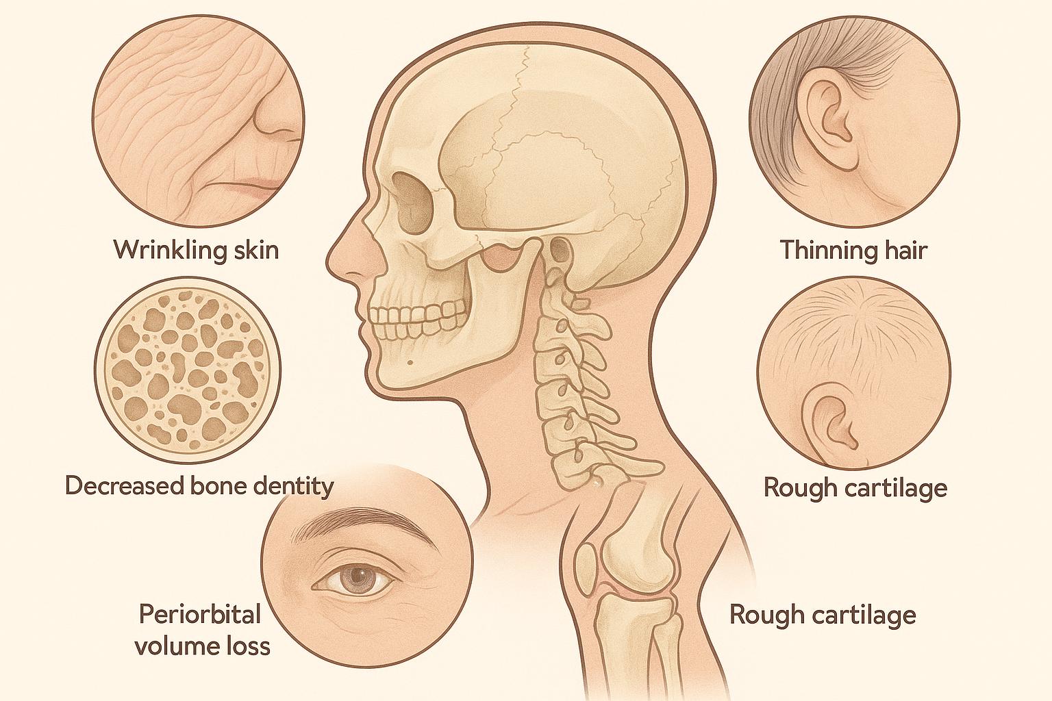
Facial aging rarely happens as a uniform drift. Instead, the structural framework of the face degrades along predictable mechanical and anatomical lines. Skin loses thickness. Fat pads shrink and shift downwards. Bone resorbs around the orbits and jawline. While superficial lines and subtle surface changes respond well to light resurfacing or soft hyaluronic acid fillers, deep structural collapse demands a completely different strategy. It requires targeted biostimulation.
True tissue regeneration relies on triggering the body’s natural extracellular matrix production.
When soft tissue loses its underlying scaffold, the visual results show up as distinct mechanical failures rather than simple superficial wrinkles. Address these structural markers with basic moisture-binding gels, and you often end up with over-filled, heavy features that lack natural contours. Biostimulatory agents like poly-L-lactic acid (PLLA) take a fundamentally different approach: they introduce micro-particles into the deep dermal or subdermal plane to trigger a controlled, sub-clinical inflammatory response that stimulates long-term type I collagen synthesis over several months.
Not all facial regions respond equally to this process. Success depends heavily on local vascularity, tissue thickness, mechanical stress, and proximity to periosteal anchorage points. Understanding which structural markers yield the highest return on investment helps clinicians avoid over-treating areas that are better served by structural implants, neurotoxins, or surgical lifting.
Table of Contents
1. Temporal Hollows and Skeletal Skeletonization
The temporal region is often the first area to show significant volume loss, yet patients rarely complain about it directly. They usually mention looking “tired,” “gaunt,” or notice that their eyebrows have lost their lateral arch.
Underneath the skin, temporal aging involves a combination of superficial temporal fat pad atrophy, deep temporal fat loss, and gradual resorption of the sphenoid and frontal bones. As these layers thin out, the lateral orbital rim becomes sharp and visible. The temporal crest stands out, creating a skeletal appearance that draws attention away from the midface.
+————————+————————————+—————————————+
| Structural Marker | Primary Anatomical Deficit | Biostimulatory Vector & Depth |
+————————+————————————+—————————————+
| Temporal Hollows | Deep/superficial fat atrophy, | Supraperiosteal / Deep fascia |
| | bone resorption at sphenoid | Depot technique with cannula or needle|
+————————+————————————+—————————————+
| Pre-Auricular Sulcus | Superficial musculoaponeurotic | Subdermal micro-droplet fanning |
| | system (SMAS) laxity, fat loss | Parallel to masseteric fascia |
+————————+————————————+—————————————+
| Midface & Zygoma | Deep medial cheek fat atrophy, | Supraperiosteal bolus + |
| | sub-orbicularis oculi fat (SOOF) | Subdermal cross-hatching overlay |
+————————+————————————+—————————————+
| Submalar Deficit | Buccal fat pad recession, | Subdermal fanning across |
| | superficial cheek fat loss | buccal area; avoid anterior movement |
+————————+————————————+—————————————+
| Pre-Jowl Sulcus | Mandibular notch resorption, | Supraperiosteal bolus anterior |
| | soft tissue descent | to mental foramen |
+————————+————————————+—————————————+
| Pyriform Aperture | Maxillary bone resorption, | Deep periosteal micro-deposits |
| | alar base recession | Medial to facial artery path |
+————————+————————————+—————————————+
Rebuilding volume in the temporal fossa requires a firm understanding of fascial layers. Injecting too superficially risks visible nodule formation, visible venous prominence, or uneven surface texture. Depositing micro-particles deeply onto the periosteum or within the deep temporal fascia allows collagen to rebuild a supportive cushion beneath the temporal muscle and superficial tissue layers.
The American Society of Plastic Surgeons highlights that deep structural pan-facial volumization provides longer-lasting, more natural facial rejuvenation than localized superficial line corrections. A restored temporal area naturally lifts the lateral brow, softens the upper orbital rim, and restores a balanced oval contour to the upper third of the face.
2. Pre-Auricular Sulcus and Lateral Cheek Flaccidity
As tissue slides forward and down, a hollow depression forms right in front of the tragus and along the ascending ramus of the mandible. This pre-auricular sulcus is a classic marker of structural aging, yet it remains under-treated in standard aesthetic plans.
When the lateral dermal matrix weakens, the entire midface soft tissue envelope loses its anchor. Think of it like a tent fabric losing tension at its outer stakes—the center sags no matter how much you prop up the middle pole.
Restoring collagen density in the pre-auricular vector tightens the lateral skin envelope. Injecting micro-particles into the subdermal plane, just above the masseteric fascia, creates a firm collagen network that pulls soft tissue back toward the ear.
Unlike hyaluronic acids—which can draw excess water and create an unnatural, puffy look along the angle of the jaw—PLLA forms dense, firm type I collagen fibers. This reinforces the lateral cheek without adding unwanted bulk or heavy width.
3. Midface Depression and SOOF Atrophy
The midface depends on a delicate stack of deep and superficial fat compartments. The sub-orbicularis oculi fat (SOOF) and deep medial cheek fat pad act as the main structural foundation for the central face.
As these deep fat pads shrink with age, the overlying skin and superficial fat lose their support. This causes the lower eyelid to blend right into the cheek, lengthening the tear trough and deepening the nasolabial fold.
Standard dermal gels can fill individual lines, but they rarely restore the broad structural platform that a youthful midface needs.
Biostimulatory collagen restoration works exceptionally well in this area because it can be placed across wide tissue planes. Injecting PLLA along the periosteum of the zygomatic arch and submalar space builds a strong scaffold under the deep fat layers.
American Academy of Dermatology clinical resources emphasize that proper patient selection and correct dilution ratios are critical when placing biostimulators near thin eyelid tissue to prevent delayed-onset nodules.
Practitioners who want to integrate these structural biostimulatory protocols into their clinical operations can buy Sculptra for aesthetic practice use to access verified injectable PLLA formulations tailored for deep dermal matrix expansion and pan-facial volume restoration. Medical clinics evaluate purchasing options based on product concentration, reconstituted stability, supply chain authenticity, and cost per vial relative to treatment course design.
Evaluating these logistical variables helps practices keep treatment costs predictable across multi-session biostimulatory protocols, which usually require two to four injection sessions spaced six weeks apart.
4. Submalar Collapse and the “Gaunt” Buccal Deficit
Submalar atrophy usually presents as a broad, hollow concavity below the zygomatic arch, stretching forward toward the corner of the mouth.
This deficit develops as the buccal fat pad recedes and superficial cheek fat thins out. It is especially prominent in athletic individuals, patients with low body fat, or those who have experienced significant weight loss.
[ Structural Aging Matrix ]
|
+————————————–+————————————–+
| |
[ Bone Resorption ] [ Soft Tissue Deficit ]| |
+–> Pyriform Recession (Nasolabial Deepening) +–> SOOF & Deep Medial Fat Atrophy
| |
+–> Mandibular Notch Erosion (Pre-Jowl Sulcus) +–> Buccal/Submalar Depletion
| |
+–> Sphenoid/Temporal Thinning (Skeletonization) +–> SMAS Laxity (Pre-Auricular Sagging)
Directly filling this wide, mobile area with soft hyaluronic gels is risky. Dynamic facial movements—like smiling or chewing—can shift the gel forward, creating a heavy, unnatural look around the corners of the mouth.
Biostimulation offers a much safer, more stable solution. By spreading a thin, cross-hatched layer of micro-particles throughout the deep submalar tissue plane, collagen fibers form evenly across the area.
This gradually thickens the dermis and subcutaneous layer without interfering with natural muscle movement. The result is a smooth, subtle transition from the cheekbone down to the jawline.
5. The Pre-Jowl Sulcus and Mandibular Notch Erosion
Jowl formation isn’t just about sagging skin; it is driven by bone loss along the jawline. Resorption of the mandibular body, combined with erosion at the pre-jowl notch, creates a distinct dip in front of the jowl fat pad.
This structural dip breaks the continuous line of the jaw, creating a wavy, aged jawline profile.
Traditional treatments often attempt to disguise the jowl by filling the depression with thick dermal gels. However, loading too much soft material along an already failing jawline can broaden the lower third of the face, making it look heavy and square.
A better strategy is to rebuild structural support directly on the bone. Placing deep, periosteal micro-deposits of PLLA at the mandibular notch stimulates localized neocollagenesis right where bone has resorbed.
This restores the underlying scaffold, smoothing the jawline without adding wide, unnatural bulk.
The American Society for Dermatologic Surgery notes that combining deep periosteal structural injections with lateral skin-tightening vectors produces significantly better jawline definition than treating the pre-jowl area in isolation.
6. Pyriform Aperture Recession and Deep Nasolabial Fold Base Collapse
The nasolabial fold is often misdiagnosed as a simple skin wrinkle. In reality, it is a structural failure caused by bone resorption at the pyriform aperture—the triangular bone opening near the base of the nose.
As the maxillary bone around the nasal pyriform aperture recedes, the alar base sinks backward into the face.
This loss of bony support drops the upper end of the nasolabial fold deep into the face, causing the overlying cheek tissue to hang over the crease.
Simply injecting soft filler directly into the crease treats the symptom, not the cause. It fills the wrinkle but leaves the underlying structural bone deficit untouched.
Rebuilding the pyriform base requires placing a deep biostimulatory foundation right on the periosteum, just lateral to the anterior nasal spine.
As new collagen forms over several months, it expands the deep tissue bed, pushing the recessed alar base forward and softening the shadow at the top of the fold naturally.
Clinical Limitations and Patient Selection Rules
Biostimulatory collagen restoration is a powerful tool, but it is not a universal fix for every patient.
Patients with severe tissue sag or hyper-lax skin rarely achieve satisfactory results from collagen induction alone. When the SMAS layer and platysma muscles have dropped significantly, surgical re-anchoring remains the gold standard.
Using biostimulators on severely lax tissue often leads to over-treatment, requiring high product volumes that yield minimal visible lift.
Proper timing and realistic expectations are essential.
Biostimulators do not offer instant gratification. Unlike hyaluronic acid gels, which provide immediate volume expansion, biostimulatory agents require time for host fibroblasts to synthesize new type I collagen matrix networks.
Patients must understand that visible improvements develop gradually over three to six months.
Furthermore, patients with active autoimmune conditions or those taking immunosuppressive therapies may produce a muted collagen response, making outcomes hard to predict.
+————————————+—————————————————+
| Clinical Variable | Diagnostic & Therapeutic Consideration |
+————————————+—————————————————+
| Severe SMAS / Skin Laxity | Surgical referral indicated; biostimulators yield |
| | poor mechanical lifting on hanging tissue. |
+————————————+—————————————————+
| Immunosuppression / Autoimmunity | Blunted fibroblast response; collagen synthesis |
| | rate unpredictable or clinically diminished. |
+————————————+—————————————————+
| Patient Expectation Timeline | Requires 3-6 months; instant volume seekers are |
| | poor candidates for biostimulatory protocols. |
+————————————+—————————————————+
| Anatomical Placement Errors | Superficial placement causes papules/nodules; |
| | strict deep subdermal/periosteal entry required. |
+————————————+—————————————————+
Nodule formation remains a known clinical risk, almost always tied to improper injection technique. Injecting too superficially, using low reconstitution volumes, or failing to massage the area properly after treatment can lead to visible or palpable PLLA clusters.
To minimize these risks, modern protocols favor higher dilution volumes, longer hydration wait times, and thorough cross-hatch fanning techniques using blunt-tip cannulas.
Strategic Integration in Structural Facial Rejuvenation
Treating facial aging successfully requires matching the right product to the specific anatomical deficit.
Superficial fine lines, lip reshaping, and sharp focal contours respond best to hyaluronic acid gels. However, broad structural hollows, bony recession, and loose lateral tissue envelopes call for targeted biostimulatory collagen restoration.
Focusing treatment on the main structural markers—the temporal hollows, pre-auricular sulcus, midface, submalar space, pre-jowl notch, and pyriform base—allows clinicians to rebuild the facial scaffold naturally.
This approach delivers subtle, long-lasting facial restoration that moves smoothly with every expression, avoiding the artificial, over-filled look of traditional superficial treatments.