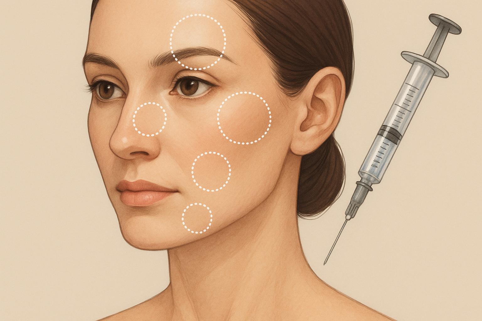
Cross-linked hyaluronic acid (HA) gels vary substantially in their viscoelastic properties. Matching the mechanical characteristics of a specific formulation—such as elastic modulus (G′), viscous modulus (G″), and tan δ—to the targeted anatomical plane determines both the longevity of the correction and the rate of adverse events. Misplacing a high-modulus, highly cohesive gel into superficial dermal layers routinely yields visible nodularity or the Tyndall effect. Conversely, placing a soft, low-G′ gel deep onto the periosteum under heavy facial musculature results in rapid displacement and inadequate structural lift.
Facial aesthetic interventions require clear mapping of tissue planes, dynamic muscle involvement, and local vascular topography. The American Society of Plastic Surgeons (ASPS) emphasizes that a thorough understanding of facial fat compartments and arterial pathways remains the primary defense against intravascular embolism and tissue necrosis.
Table of Contents
1. Deep Malar and Sub-Orbicularis Oculi Fat (SOOF) Planes
Midface volumetric loss occurs secondary to both deep fat pad atrophy and maxilla resorption. Restoring projection in the cheek complex demands a gel capable of withstanding significant shear stress from the zygomaticus major and minor muscles.
High G′ formulations function as structural surrogates when injected supraperiosteally in the midface. In these deep planes, the tissue requires maximum lifting capacity rather than high flexibility.
MIDFACE LAYERS
━━━━━━━━━━━━━━━━━━━
│ Skin │
├━━━━━━━━━━━━━━━━━━━┤
│ Subcutaneous │
├━━━━━━━━━━━━━━━━━━━┤
│ SMAS / Muscle │
├━━━━━━━━━━━━━━━━━━━┤
│ Deep Fat (SOOF) │
├━━━━━━━━━━━━━━━━━━━┤
│ Periosteum/Bone │ ◄── Place High-G’ Gel Here
└━━━━━━━━━━━━━━━━━━━┘
When targeting the deep malar space, clinicians typically employ a bolus technique directly on the bone. The soft tissue envelope above the gel must be thick enough to obscure the product’s boundaries.
2. The Pyriform Aperture and Nasolabial Fold
The nasolabial fold represents a combined problem of superficial skin creasing and deep structural deflation at the pyriform aperture. The facial artery runs in close proximity to this region, altering its depth as it travels superiorly toward the alar base.
Injecting the upper aspect of the nasolabial fold carries elevated risk. The American Society for Dermatologic Surgery (ASDS) clinical guidelines highlight the alar base as a high-risk zone for vascular compromise due to branches of the angular artery.
NASOLABIAL FOLD: ANATOMICAL STRATIFICATION & GEL REQUIREMENTS
Superficial Dermis ──► [ Flexible, Moderate-G’ HA Gel ]
(Dynamic movement; prevents ridging)
│
Subcutaneous Space ──► [ Intermediate Zone – Caution: Vessels ]
│
Pyriform Aperture ──► [ High-G’, High-Cohesivity Gel ]
(Supraperiosteal bolus for deep support)
Deep structural loss at the pyriform aperture is corrected via supraperiosteal placement of high-density gels. Finer superficial lines within the fold, however, require lower G′ formulations administered intradermally or into the immediate sub-dermal plane.
3. The Mandibular Angle and Line
Lower face rejuvenation often involves re-establishing definition along the jawline. Age-related bone resorption at the mandibular angle, combined with jowl formation, obscures the structural transition between the neck and the lower third of the face.
Recreating a sharp mandibular angle requires a firm gel with high resistance to deformation.
JAWLINE TARGET ZONES
Mandibular Angle
(Deep Supraperiosteal) ──► High G’ / Firm Structural Gel
│
▼
Pre-Jowl Sulcus
(Subcutaneous) ──► Moderate G’ / Cohesive Gel
For medical practices sourcing specialized dermal matrices for these varied structural depths, securing predictable product batches remains a core operational requirement. Licensed practices often rely on a Stylage supplier for professional orders to maintain inventory of varying cross-linking densities, ranging from light formulations for superficial fine lines to denser gels designed for periosteal structural augmentation. Having access to full product ranges allows clinicians to match the exact rheological profile to the targeted tissue layer.
Product density selections directly dictate needle gauge requirements and extrusion force, factors that influence precise delivery in high-density tissue spaces like the chin and ramus.
4. The Temporalis Fascia and Deep Temporal Fossa
Temporal wasting creates a skeletonized upper facial appearance. The temporal region contains complex fascial layers, including the superficial temporal fascia, the deep temporal fascia, and the temporalis muscle.
TEMPORAL LAYERS
━━━━━━━━━━━━━━━━━━━━━━┓
│ Skin │
├━━━━━━━━━━━━━━━━━━━━━┤
│ Superficial Temporal Fascia (STF)
│ ── Contains Superficial Temporal Artery
├━━━━━━━━━━━━━━━━━━━━━┤
│ Loose Areolar Tissue
├━━━━━━━━━━━━━━━━━━━━━┤
│ Deep Temporal Fascia
├━━━━━━━━━━━━━━━━━━━━━┤
│ Temporalis Muscle
├━━━━━━━━━━━━━━━━━━━━━┤
│ Periosteum / Bone│ ◄── Injection Target Zone
└━━━━━━━━━━━━━━━━━━━━━┘
Deep injection onto the periosteum beneath the temporalis muscle isolates the gel from superficial movement and minimizes the visibility of product edges. Medium-to-high G′ gels spread predictably in this space when gentle post-injection massage is applied.
5. The Vermilion Border and Lip Parenchyma
The lips present a distinct mechanical environment characterized by constant high-frequency dynamic movement from the orbicularis oris muscle. Gels placed in the lip body must possess high stretchability and moderate cohesivity to prevent lumps or artificial resistance during speech and mastication.
LIP ANATOMY & TARGET PLANES
Vermilion Border ──► Precise linear threading (Low/Moderate G’)
Restores border definition.
Dry Vermilion ──► Depot / Volumizing bolus (High Stretchability)
Restores mucosal volume.
High G′ gels are generally contraindicated in the lip parenchyma because they feel unnaturally firm to the patient and tend to migrate under continuous muscular shear. Moderate-density gels optimized for dynamic elasticity yield smoother integration within the submucosal layer.
6. The Dorsum and Nasal Tip (Non-Surgical Rhinoplasty)
The nasal dorsum consists of an osteocartilaginous framework covered by a thin soft tissue envelope. Because the tissue space is restricted and vascularization relies on the dorsal nasal and lateral nasal arteries, non-surgical rhinoplasty demands extreme caution and precise gel selection.
High-cohesivity, high-modulus gels are preferred here to prevent lateral spreading down the nasal sidewalls.
NASAL DORSUM INJECTION PLANE
Skin / Subcutaneous Layer
─────────────────────────────
SMAS & Vascular Plexus ◄── AVOID
─────────────────────────────
Deep Submuscular Plane ◄── TARGET ZONE
─────────────────────────────
Periosteum / Cartilage
Injecting strictly in the midline on the perichondrium or periosteum lowers the risk of vascular cannulation and keeps the gel stable under skin tension.
Rheological Matrix Comparison
| Anatomical Zone | Primary Tissue Plane | Target Gel Modulus (G′) | Primary Rheological Requirement | Key Risk Factor |
| Midface / Malar | Supraperiosteal / Deep SOOF | High | High lifting capacity | Transverse facial artery branches |
| Pyriform Aperture | Deep Supraperiosteal | High | Structural projection | Angular artery occlusion |
| Mandibular Angle | Periosteal | Very High | High deformation resistance | Facial artery at the facial notch |
| Temporal Fossa | Periosteal (Deep to muscle) | Medium to High | Spreadability under muscle | Sentinel vein & deep temporal arteries |
| Lip Parenchyma | Submucosal | Low to Medium | High stretchability (tanδ) | Superior/Inferior labial arteries |
| Nasal Dorsum | Deep Perichondrial/Periosteal | High | High cohesivity (no spreading) | Dorsal nasal artery & lateral necrosis |
Clinical Limitations and Safety Considerations
While hyaluronic acid gels offer versatility due to their reversibility with hyaluronidase, several clinical limitations exist across all anatomical zones:
- Vascular Occlusion: Inadvertent intra-arterial injection can lead to immediate ischemia, skin necrosis, or vision loss. Knowledge of local anatomy does not eliminate risk due to anatomical variations among patients.
- Late-Onset Nodule Formation: Delayed-type hypersensitivity reactions or biofilm formation can occur months post-injection, particularly with highly cross-linked, high-density gels.
- Product Migration: Incorrect gel selection (e.g., placing a low-cohesivity gel in a high-muscle-activity zone) often leads to product displacement over time.
- Tissue Compression: Excessive volume placement in tight anatomical compartments (such as the nasal tip) can cause extrinsic vascular compression without direct intra-arterial entry.
Adhering strictly to plane-specific rheological selection criteria minimizes mechanical failures and optimizes clinical outcomes across all facial regions.